
 Türkçe
 Deutsch
 Français

Reconstructive approaches, functional hearing restoration, and minimally invasive methods in tympanoplasty surgery involve the anatomical reconstruction of the ruptured eardrum and damaged ossicular chain without any externally visible incision, using transcanal endoscopic systems and durable autologous cartilage micro-grafts. This modern surgical approach proceeds through the natural opening of the external ear canal, clears the focus of infection in the middle ear, and repairs the sound transmission mechanism in the same session, providing the patient with a safe, water-resistant ear structure with restored hearing function. Micro-reconstruction techniques that fully preserve the morphology of the auricle and surrounding tissues provide permanent tissue stability and eliminate the need for invasive open surgery.
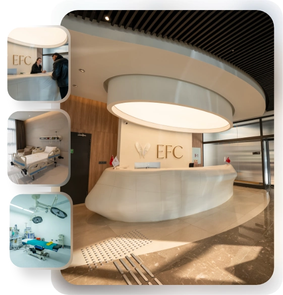
EFC Surgery Center
Comprehensive Care: From Initial Consultation to Follow-Up.
EFC Surgical Medical Center is a center of excellence specializing in the most meticulous fields of surgical medicine, from aesthetic surgery to interventional treatments—where every step progresses with refined attention. Medical excellence, aesthetic precision, and uncompromising ethical standards converge on the same path. Our subspecialty-trained experts aim to achieve natural and reliable results by delivering evidence-based care supported by modern imaging, standardized protocols, and safety systems. From consultation to recovery, your care is coordinated end-to-end with clear communication, transparent planning, and genuine respect for your health.
What is tympanoplasty (eardrum surgery) and in which situations is it generally planned?
The eardrum is a highly sensitive structure that collects incoming sound waves, vibrates, and transfers these vibrations to the tiny ossicular chain in the middle ear. A healthy eardrum should maintain its integrity and isolate the middle ear from the external environment. However, conditions such as frequent ear infections during childhood, severe physical trauma to the ear, and exposure to loud sound or pressure can cause tears in the eardrum. Although small tears often tend to heal on their own, these perforations can become permanent, especially in cases accompanied by chronic ear infections.
A perforated eardrum means that the middle ear is left completely vulnerable to water, dust, and various bacteria coming from the outside world. This condition creates a basis for recurrent ear discharge that significantly reduces the quality of daily life. The main purpose of the tympanoplasty procedure is to break this cycle of chronic inflammation, close the ear safely and healthily, and also minimize the patient’s concerns regarding contact with water. The surgery aims to ensure that the middle ear and the surrounding air spaces are covered with healthy mucosa.
How does a perforation in the eardrum affect the hearing process and what are the symptoms that may require tympanoplasty (eardrum surgery)?
A healthy hearing process occurs when sound waves strike the eardrum and these vibrations are transmitted to the inner ear through the malleus, incus, and stapes in sequence. You can think of the eardrum as a tightly stretched drumhead. When there is a perforation in the eardrum, sound waves leak inward through this opening instead of accumulating on the surface of the membrane and increasing their strength. This mechanical impairment disrupts the natural amplification stage of sound and causes hearing loss commonly known as “conductive” hearing loss.
Depending on the size of the perforation and its location on the eardrum, individuals may have difficulty understanding everyday conversations or may feel the need to turn up the television volume more than usual. Untreated and constantly recurring infections may, over time, impair the nourishment of the delicate hearing ossicles inside and cause them to erode. A process that initially begins as a simple perforation in the eardrum has the potential to develop over the years into a much more complex condition in which the ossicles are damaged.
Some of the main complaints that may require tympanoplasty surgery are as follows:
- Hearing loss
- Ear discharge
- Buzzing
- Ringing
- A feeling of fullness in the ear
- Recurrent infections
How does the process before tympanoplasty (eardrum surgery) proceed and which tests are requested from the patient for surgical preparation?
One of the most important steps of successful eardrum surgery is ensuring that the ear to be operated on is free of infection and dry before the operation. In operations performed during a period when the ear is inflamed, actively discharging, and the tissues are swollen, the grafts used for repair generally have a lower chance of taking. For this reason, the preoperative process usually includes the stage of drying the ear with medical treatments and ear drops.
During the clinical evaluation stage, the eardrum is examined in great detail with the help of a microscope or endoscope. The size of the perforation, the structure of its edges, and the health of the middle ear tissue are evaluated. Hearing tests are performed to understand how much of the hearing loss is caused by the perforation in the eardrum and how much is caused by a possible ossicular or inner ear problem. Radiological imaging methods are used especially if the disease is suspected of having damaged the ossicles or if the patient has previously undergone ear surgery. In this way, the deep recesses of the middle ear and the general condition of the ossicles are mapped before surgery.
Some of the examinations that may be requested from the patient during surgical planning can be listed as follows:
- Pure-tone audiometry
- Tympanometry
- Computed tomography
- Routine blood tests
What are the types of tympanoplasty (eardrum surgery) and how is surgery classified according to the degree of damage in the middle ear?
The level of damage in each patient’s ear structure is different; therefore, eardrum surgeries cannot be considered a standard, single-type procedure. In the medical approach, the scope of surgery is determined according to the level of damage in the middle ear and is divided into various types.
If there is no problem with the hearing ossicles and the problem consists only of a perforation in the eardrum, the procedure is performed simply by patching this perforation. However, if the inflammation has damaged the malleus, which is the first link in the ossicular chain, or the incus, which is the second link, the tissue used for repair is brought into direct contact with the remaining intact ossicles in an attempt to re-establish the sound transmission bridge.
In more advanced damage, both the malleus and incus may have lost their function. In this scenario, only the stapes, the last ossicle that acts as the gateway to the inner ear, remains. In this case, specially designed prostheses or shaped cartilage pieces are used to transmit sound waves directly to the stapes or its base. The method to be selected according to the extent of the disease is decided in light of the preoperative tests and the findings during surgery.
Contact us now to get detailed information about our treatments and procedures and to schedule an appointment!
What is endoscopic tympanoplasty (eardrum surgery) and how can the innovations it offers compared with traditional open surgeries be summarized?
In the past, eardrum repairs were generally performed through large incisions made behind the auricle. Although this traditional method provided a wide working area, it had some disadvantages such as leaving a scar behind the ear, requiring dressings during the recovery period, and causing greater discomfort for the patient. There was also the possibility of an asymmetrical appearance of the auricle after the operation.
With the development of modern medicine and optical systems, “Transcanal Endoscopic Ear Surgery” has begun to come to the forefront. In the endoscopic method, the surgery is performed entirely through the natural opening of the external ear canal. Thanks to thin endoscopes with special angled lenses, the most hidden corners of the middle ear, which traditional microscopes have difficulty illuminating with their straight viewing angle, can be observed on high-resolution screens. In this way, there is generally no need to drill healthy bone tissues solely to obtain a viewing angle, and an attempt is made to preserve the natural anatomy of the ear.
What are the aesthetic advantages offered to patients by the incisionless endoscopic tympanoplasty (eardrum surgery) approach?
The integration of aesthetic surgery principles into ear operations has provided significant improvements in patient comfort. The greatest advantage of the endoscopic method for patients is that it aims not to cause any change in external appearance. Since no large incision is made behind the ear, tissue trauma is minimized and procedures such as suture removal are generally not required.
The application of the incisionless method also positively affects the level of pain experienced by patients after surgery. Compared with traditional methods, a large proportion of patients who undergo endoscopic surgery can return to their normal lives much faster. Since the need for head bandages or heavy dressings lasting for weeks is eliminated, patients can go through the process much more comfortably.
The aesthetic and functional advantages that the endoscopic method is generally considered to provide to the patient are as follows:
- No externally visible incision
- Preservation of the natural ear angle
- Lower pain profile
- Faster recovery process
- Possibility of same-day discharge
What tissue grafts are used in tympanoplasty (eardrum surgery) repair and why is cartilage use preferred more often?
To close the perforation in the eardrum, natural tissues taken from the patient’s own body (autologous grafts) are always used instead of artificial materials. For many years, the “fascia” tissue, which is the thin membrane of the muscle above the ear, was used as the standard for this purpose. Because fascia is thin and flexible, it adapts to sound transmission. However, because it has a weak blood supply, it carries the risk of dissolving over time or collapsing inward because it cannot withstand the negative pressure in the middle ear.
In current ear surgery, the use of cartilage tissue has largely changed the standards. A thin cartilage tissue taken from the patient’s own ear has a highly resistant structure that requires very little nourishment. Cartilage is more resistant to infections and prevents collapse of the membrane by resisting the negative vacuum effect that may occur in the middle ear. Cartilage membranes shaped to an appropriate thinness aim to provide a much longer-lasting anatomical structure while being able to transmit sound as well as flexible muscle fascia.
Does the cartilage taken for eardrum surgery (tympanoplasty) affect external appearance and which cartilage shaping methods are used?
Cartilage tissue is generally obtained from the small prominence just in front of the ear (tragus) or from the bowl-shaped depression of the auricle. At this point, patients usually wonder whether the shape of their ears will be distorted. In modern approaches where the rules of facial aesthetics are meticulously observed, the natural form of the ear is preserved. When cartilage is taken from the tragus region, the incision is hidden on the inner surface of the ear and is intended not to leave a visible scar from the outside. Most importantly, the entire cartilage is not removed; the edge portion of the cartilage framework that defines the outer contours is left in place, thereby preventing any collapse or asymmetry.
The cartilage obtained is shaped using different techniques depending on the size of the perforation in the membrane. For small and medium-sized perforations, the method called “butterfly cartilage” is frequently used. In this technique, the cartilage is grooved along its edges, much like a spool or a wick, and locked into the perforation. For larger perforations or completely collapsed membranes, the “palisade” (fence) technique is preferred. In this method, the cartilage is cut into long, thin strips and placed side by side beneath the perforation, thereby both ensuring nourishment of the tissue and creating a strong wall.
When the middle ear ossicles have eroded, which procedures are performed during tympanoplasty (eardrum surgery) to improve hearing?
Chronic ear infections can cause structural erosion of the hearing ossicles in the middle ear over the years. When the connection between the malleus and the stapes is disrupted, sound cannot reach the inner ear no matter how well the eardrum vibrates. The procedure performed to re-establish this mechanical connection of the hearing system is called “ossiculoplasty.”
If the discontinuity or gap between the ossicles is very small, special biocompatible bone cements can be used to bridge this gap. Bone cement improves the connection by providing a natural vibration when it hardens. However, if a significant portion of the ossicles has eroded, special micro-prostheses made of titanium or bone-like materials are used to fill the distance between them. When these prostheses are placed, a thin cartilage cushion is always positioned between the head of the prosthesis and the eardrum in order to prevent the metal from pressing on the new membrane over time and perforating it. In this way, sound is expected to be distributed more naturally across the membrane.
Contact us now to get detailed information about our treatments and procedures and to schedule an appointment!
What is the condition called cholesteatoma and what is its general relationship with tympanoplasty (eardrum surgery)?
One of the most serious forms of chronic middle ear disorders is the condition called cholesteatoma. In simple terms, cholesteatoma is the unwanted progression of skin cells belonging to the eardrum into the middle ear and the air-filled bone spaces within the skull, where they form cyst-like structures. As these skin cells grow, they have the potential to erode the healthy bone tissue around them.
Although cholesteatoma is not a cancer or tumor, due to its progressive nature it can destroy the hearing ossicles, threaten the facial nerve, or, in very advanced stages, create a basis for intracranial problems. In these cases, the absolute priority of surgery is to completely clear the patient of this destructive tissue rather than to correct the impaired hearing. During surgery, the diseased tissue is meticulously removed. Depending on the extent of the disease, the procedure is performed either by preserving the posterior bony wall or by opening it completely. The use of endoscopic systems is an important aid in reducing the risk of recurrence of the disease because it allows residual tissue in inaccessible blind spots to be seen. Reconstruction of hearing is generally planned in stages after the ear has been made safe.
How does the recovery process proceed after tympanoplasty (eardrum surgery) and what important rules should patients follow?
After the surgery is completed, special self-dissolving gel sponges may be placed in the external ear canal to support the newly created eardrum and help it heal in the correct position. During the first weeks, hearing may feel somewhat muffled or reduced due to the presence of these sponges and postoperative swelling in the ear, and this is considered a natural part of the healing process.
The first few weeks are a very critical period because the new eardrum has not yet reached its full strength. Patients are expected to strictly avoid movements that could create sudden pressure changes in the middle ear during this period. Completely isolating the ear from water is another vital rule for the graft to heal healthily without becoming infected. It is recommended to place suitable protectors at the entrance of the external ear canal while bathing. It is generally expected that within a few months the membrane will fully settle into place and the hearing level will reach its final form.
The main rules that patients are advised to follow in the early postoperative period are as follows:
- Protecting the ear from water
- Not blowing the nose
- Keeping the mouth open while sneezing
- Not lifting heavy objects
- Avoiding constipation
As with every surgical intervention, what are the possible risks and complications associated with tympanoplasty (eardrum surgery) procedures?
Although success rates have reached very high levels with the use of modern medical devices and advanced surgical techniques, it should not be forgotten that every surgical intervention carries certain risks by its nature. One of the most common situations that may be encountered is that the cartilage or fascia graft placed does not fully adapt to the tissue and a perforation forms again in the eardrum. This condition is generally associated with a new upper respiratory tract infection occurring in the early postoperative period or with the patient’s pressure equalization mechanism between the nose and ear not functioning properly.
A thin nerve that carries the sense of taste passes very close to the surgical area. Depending on the procedures performed during surgery, some patients may experience temporary metallic taste changes or taste disturbances on one half of the tongue; this condition generally tends to resolve spontaneously within months. In much rarer cases, conditions such as incorrect healing of the eardrum, dizziness due to involvement of the inner ear, or persistence of hearing problems may occur. More serious risks such as facial paralysis have become very rare thanks to special nerve monitoring devices used in modern surgeries.
Possible risks that may be encountered, although rarely, in tympanoplasty surgery are as follows:
- Failure of the graft to take
- Temporary taste disturbance
- Persistence of hearing loss
- Dizziness
- Collapse of the eardrum
Blog Posts

Hair Transplant Prices
Hair transplant prices vary depending on the technique to be used, the number of grafts, [...]
Read More
21
Jan
Jan

Most Preferred Countries for Plastic Surgery
Among the most preferred countries for plastic surgery, Turkey, South Korea, the United States, and [...]
Read More
21
Jan
Jan

Plastic Surgery Prices
Plastic surgery prices vary depending on the type of procedure, geographical location, the surgeon’s experience, [...]
Read More
21
Jan
Jan

Physical Therapy Prices
Physical therapy prices vary depending on the number of sessions to be applied, the treatment [...]
Read More
21
Jan
Jan

Dental Treatment Prices
Dental treatment prices vary depending on the type of procedure to be performed, the quality [...]
Read More
21
Jan
Jan

Most Preferred Countries for Dental Treatments
Among the most preferred countries for dental treatments are Turkey, Hungary, Mexico, and Thailand. These [...]
Read More
21
Jan
Jan