
 Türkçe
 Deutsch
 Français

Endometriosis surgery is the surgical removal of lesions, chocolate cysts, and pelvic adhesions caused by tissue resembling the inner lining of the uterus growing outside the uterus. This operation aims to repair the physical damage caused by the disease in surrounding organs and restore the pelvic anatomy to its natural form. It is based on removing not only superficial lesions but also hardened nodules that have infiltrated deep tissues according to the principle of excision, while preserving the healthy structures around them. Designed to eliminate the mechanical tension caused by this progressive disease, the surgical process focuses on supporting the normal functioning of the organs and restoring balance to the disrupted dynamics of the body.
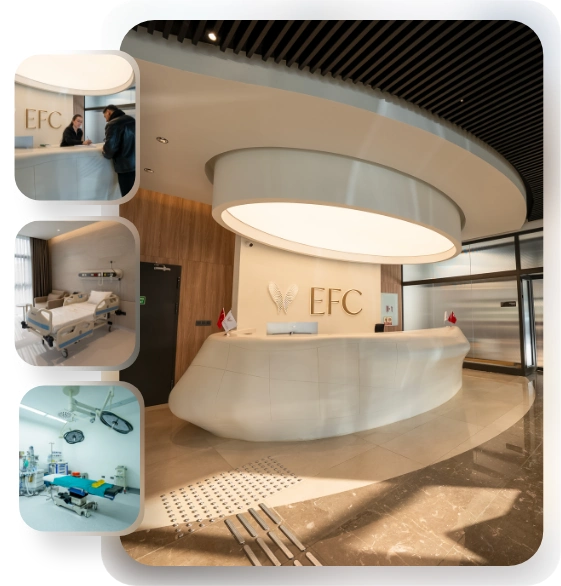
EFC Surgery Center
Comprehensive Care: From Initial Consultation to Follow-Up.
EFC Surgical Medical Center is a center of excellence specializing in the most meticulous fields of surgical medicine, from aesthetic surgery to interventional treatments—where every step progresses with refined attention. Medical excellence, aesthetic precision, and uncompromising ethical standards converge on the same path. Our subspecialty-trained experts aim to achieve natural and reliable results by delivering evidence-based care supported by modern imaging, standardized protocols, and safety systems. From consultation to recovery, your care is coordinated end-to-end with clear communication, transparent planning, and genuine respect for your health.
What is endometriosis surgery?
There is a special tissue that forms the inner lining of the uterus, and this tissue thickens every month in preparation for pregnancy; if pregnancy does not occur, it is expelled from the body through menstrual bleeding. When these cells settle outside the uterus, for example on the ovaries, pelvic membranes, or intestines, and continue to live there, endometriosis develops. These tissues scattered in different parts of the body attempt to bleed every month under the influence of estrogen, the female hormone, just as if they were inside the uterus. However, there is no pathway for this blood to leave the body. The blood trapped inside initiates chronic inflammation in the surrounding tissues. Over time, the inflamed areas heal and contract, turning into very hard scar tissue and thick adhesions that bind organs together. Since medication cannot eliminate these mechanical adhesions and hardened nodules, surgical intervention is required to restore the anatomy.
Which symptoms do patients most commonly experience before the decision for endometriosis surgery is made?
As the disease progresses, the inflammation and adhesions it causes in the body begin to produce very pronounced signs that seriously reduce quality of life. In particular, cramps that intensify during menstrual periods and do not ease even with painkillers can bring women’s lives almost to a standstill. In addition, lesions located on the ligaments behind the uterus or deep in the vagina can make sexual life very difficult. When the disease progresses to surrounding organs, even bowel movements can become painful.
Common clinical complaints include:
- Chronic pelvic pain
- Severe menstrual pain
- Painful sexual intercourse
- Painful bowel movements
- Unexplained infertility
How is preoperative mapping performed for a successful endometriosis surgery?
For surgery to proceed efficiently and for possible surprises to be avoided, the extent of the disease in the body must be mapped in great detail before the operation. Each patient’s anatomy and disease course are unique, just like a fingerprint. With a detailed gynecological examination, hard areas behind the uterus, possible nodules in the vaginal wall, and the mobility of the pelvic organs are evaluated. Advanced ultrasonography systems are used to check whether the uterus and intestines naturally slide over one another. If there is no sliding movement, it is understood that the organs are densely adhered to each other, and the surgical plan is revised to be more comprehensive in light of this information.
How do imaging techniques guide the planning of endometriosis surgery?
Especially in cases where the disease is suspected of infiltrating deep tissues, examination and standard ultrasound alone may not be sufficient. At this point, magnetic resonance imaging (MRI) with special imaging protocols is used to visualize the boundaries of the disease at the millimetric level. In this way, it can be seen which layer of the intestinal wall the disease has reached, whether it is causing compression in the urinary tract, and to what extent it involves the ligaments around the uterus. Before entering the operating room, the surgical team determines which organs will require intervention and to what extent based on these images.
The regions examined in detail on MRI include:
- Ovaries
- Ligaments behind the uterus
- Colon wall
- Urinary bladder
- Ureteral canals
Why is endometriosis surgery preferred when treatment with medication alone is possible?
Medical treatments, especially hormone-suppressing pills or intrauterine devices, help relieve pain by reducing the estrogen support on which the disease depends. However, these medications only put the disease into a dormant state; they cannot physically dissolve hardened nodules that have formed over the years, fibrous bands that bind organs together, or enlarged chocolate cysts. If pain continues despite medication, or if the disease has reached a level that threatens kidney or intestinal functions, surgery ceases to be merely an alternative and becomes a fundamental necessity. The operation aims to eliminate mechanical tension in the tissues.
Why is minimally invasive endometriosis surgery performed more often than open surgery in modern medicine?
In previous years, traditional surgical methods involving a large incision in the abdomen were used. However, in open surgery, the abdominal organs come into contact with air, causing tissue drying; this can predispose the organs to adhere to each other more during the healing period. In addition, it is very difficult to illuminate the deepest and darkest areas of the pelvis with the naked eye. Today, minimally invasive methods are preferred. In closed operations, the abdomen is gently inflated with carbon dioxide gas to create a wide working space, and thanks to high-resolution cameras, even the finest blood vessels and nerves are projected onto the screen at a greatly magnified size. This allows less damage to be caused to the tissues.
Contact us now to get detailed information about our treatments and procedures and to schedule an appointment!
How is laparoscopic endometriosis surgery performed step by step?
Laparoscopy is performed using special instruments inserted into the abdomen through several millimetric incisions made at the belly button and groin areas. The optical camera introduced inside allows millimetric disease lesions that are very difficult to see with the naked eye to be easily distinguished. To separate the tissues from each other, the surgeon generally uses a gentle separation technique called hydrodissection, which makes use of water pressure. Diseased areas are carefully stripped from the surrounding membranes, much like separating the core of an onion, and the diseased tissues are removed through small incisions.
The main surgical instruments used during the operation include:
- High-resolution cameras
- Cold light sources
- Energy devices for controlling bleeding
- Atraumatic graspers
- Fine dissection scissors
What advantages do advanced robotic systems provide during endometriosis surgery?
The robotic platform takes all the advantages offered by laparoscopy to a higher technological level. The surgeon sits at a special console beside the patient and manages the operation with three-dimensional imaging that provides true depth perception. The robot’s arms are not subject to the natural limitations of the human wrist; they can rotate at much wider angles, making suturing easier even in the narrowest pelvic spaces. They also filter out and eliminate even the slightest natural tremors of the human hand. In cases where the organs are adhered to each other like concrete, this high degree of maneuverability and precision significantly increases surgical comfort.
How is endometriosis surgery performed in chocolate cyst cases without damaging the ovary?
Endometriomas, commonly known as chocolate cysts, are not actually true cysts with their own independent capsule. They form when diseased tissue on the outer surface of the ovary collapses inward and bleeds during each menstrual period, causing the tissue to swell like a balloon. The false membrane that forms around the cyst adheres very tightly to the tissue that contains the woman’s healthy eggs. The most delicate stage of surgery is avoiding the accidental removal of healthy ovarian tissue together with the cyst while peeling it away. Finding the correct anatomical plane and separating the tissues with fine movements helps preserve ovarian capacity.
How is ovarian reserve, which determines women’s fertility, affected after endometriosis surgery?
Every surgical procedure has a certain effect on tissues. After the cyst is removed, microbleeding naturally begins in the remaining ovarian bed. Using intense high heat to burn the tissue in order to stop this bleeding (cauterization) can impair the microscopic blood circulation of the ovary and cause a permanent reduction in reserve. To prevent this, the use of heat is avoided as much as possible. Bleeding is controlled using biological adhesives, special sponges, or extremely fine absorbable sutures, and preservation of the reserve is largely targeted.
How is the endometriosis surgery plan personalized in women whose ovarian reserve is already low?
In women who have previously undergone ovarian surgery, have giant cysts in both ovaries, or are found to have a significantly reduced egg pool in preoperative blood tests, standard cyst stripping may pose a risk of early menopause. In this special patient group, alternative approaches are adopted. Instead of completely stripping the cyst wall, the thick dark fluid inside is drained and cleaned.
Alternative steps used in patients with low reserve include:
- Intracystic drainage
- Superficial vaporization
- Egg freezing
- Embryo freezing
Why is surgery for deeply infiltrating endometriosis with extensive disease a more challenging process?
When the disease extends several millimeters below the membrane lining the abdominal cavity and infiltrates the muscle tissue of surrounding organs, it is called deep infiltrating endometriosis. This is one of the most difficult stages of gynecological surgery and requires advanced knowledge of anatomy. The intense inflammatory reaction causes the uterus, intestines, and urinary tract to become fused into a single mass. The boundaries between healthy tissue and diseased tissue become indistinct. In such difficult cases, the operation is generally carried out jointly by a broad surgical team that also includes general surgery and urology specialists.
Which approaches are followed during endometriosis surgery when the lesions reach the intestines?
When deep nodules settle in the final section of the large intestine, pain during bowel movements and signs of intestinal obstruction begin to occur. The basic philosophy here is to remove the lesion while preserving intestinal integrity. If the nodule is small and superficial, only the outer surface is shaved away as a thin layer. If the disease has penetrated deeper, the affected area is removed as a round piece approximately the size of a coin and the resulting defect is quickly sutured. However, if the inner lumen of the intestine is largely obstructed, the diseased segment may need to be completely cut out and the remaining healthy ends joined together.
Techniques used in intestinal surgery include:
- Superficial shaving
- Discoid resection
- Segmental resection
- End-to-end anastomosis
Contact us now to get detailed information about our treatments and procedures and to schedule an appointment!
How does endometriosis surgery protect the kidneys when the urinary tract is at risk?
The urinary tract is one of the main regions where the disease can progress silently. The ureteral canals that carry urine from the kidneys may become narrowed due to pressure from surrounding hard nodules. Even if the patient feels no pain, urine can flow backward and cause kidney enlargement and loss of function. During surgery, the ureter is first freed by releasing it from surrounding adhesions. If the structure of the canal is damaged, that section is removed and the healthy portion is connected to the urinary bladder through a new pathway. Lesions infiltrating the urinary bladder are removed by cutting within healthy tissue margins, and the bladder is repaired with special techniques in accordance with its original form.
How does endometriosis surgery performed while preserving autonomic nerves support a return to normal life?
The inside of the pelvis is covered with a very fine, spiderweb-like nerve network that enables the sensation of needing to use the toilet, emptying of the bladder, and stimulation of sexual functions. Hard endometriosis nodules generally develop around or directly over these nerves. If these invisible nerve fibers are cut or damaged by heat while clearing the disease, patients may experience prolonged inability to urinate or permanent constipation after surgery. For this reason, surgical dissection procedures are performed with special maneuvers specifically aimed at protecting these nerves, and nerve damage is minimized as much as possible.
How does the body’s recovery process progress after extensive endometriosis surgery?
The greatest advantage of minimally invasive surgical techniques for the patient is that recovery is quite rapid. After operations in which only a cyst is removed or superficial lesions are cleared, patients can usually leave the hospital within one or two days and return to their normal working lives within a few weeks. In more complex procedures involving intestinal or urinary tract repairs, the biological healing process of the tissues naturally takes somewhat longer. In these patients, normalization of bowel movements and healing of internal sutures require a few weeks of rest.
The main recommendations supporting recovery include:
- Early mobilization
- Plenty of fluid intake
- Short walks
- Fiber-rich nutrition
- Regular rest
Is there a possibility that the disease may recur even after a very well-performed endometriosis surgery?
Because the disease is associated with the immune system and genetic predisposition, there may be a risk of recurrence no matter how comprehensive the surgery is. While the rate of recurrence is higher when superficial lesions are merely burned and left in place, complete excision of diseased tissues together with the surrounding reactive area helps significantly reduce the risk of recurrence. However, as long as a woman continues to menstruate and produce hormones, microscopic cells lying dormant elsewhere in the body have the potential to become active over time.
Which supportive treatments are used after endometriosis surgery to suppress the risk of recurrence?
After all visible lesions causing symptoms are removed from the body through surgery, a medical protection plan is introduced to stop the growth of any remaining lesions at the cellular level. If the patient does not plan to have a baby in the near future, special hormone medications or intrauterine devices that balance estrogen levels and reduce menstrual activity are used. This medical suppression process largely helps prevent the disease from flaring up again by effectively allowing the pelvic region to rest.
How long should women who want to become mothers in the future wait for pregnancy after endometriosis surgery?
The chronic inflammation caused by the disease around the uterus can make it difficult for sperm and egg to meet. After surgery, when the tubes and ovaries are restored to their natural positions and the inflammatory burden is removed from the body, the reproductive system can function more freely. The first one-year period following surgery is called the “golden window.” Since anatomical obstacles have been removed during this period, the couple’s chance of achieving spontaneous pregnancy is at its highest. If there is no additional problem, couples are generally advised to try naturally during this period.
At what stage should endometriosis surgery be considered in couples planning IVF treatment?
Since every patient’s reproductive history is different, the sequence of surgery and IVF treatment must be planned entirely on an individual basis. If the woman is young and her ovarian reserve is sufficient, surgery may be performed first to overcome anatomical obstacles. However, if the woman’s age is advanced and a serious reduction in ovarian capacity has been identified, stimulating the remaining eggs and freezing embryos before surgery may be preferred.
The main factors taken into consideration include:
- Woman’s age
- Ovarian reserve
- Condition of the fallopian tubes
- Sperm quality
In this way, after the patient’s reproductive potential is secured, surgery is planned with the aim of preparing a clean and pain-free anatomical environment in which the embryo can implant. Endometriosis surgery provides valuable support for women’s health when performed at the right time and with the correct techniques.
Blog Posts

Hair Transplant Prices
Hair transplant prices vary depending on the technique to be used, the number of grafts, [...]
Read More
21
Jan
Jan

Most Preferred Countries for Plastic Surgery
Among the most preferred countries for plastic surgery, Turkey, South Korea, the United States, and [...]
Read More
21
Jan
Jan

Plastic Surgery Prices
Plastic surgery prices vary depending on the type of procedure, geographical location, the surgeon’s experience, [...]
Read More
21
Jan
Jan

Physical Therapy Prices
Physical therapy prices vary depending on the number of sessions to be applied, the treatment [...]
Read More
21
Jan
Jan

Dental Treatment Prices
Dental treatment prices vary depending on the type of procedure to be performed, the quality [...]
Read More
21
Jan
Jan
Most Preferred Countries for Dental Treatments
Among the most preferred countries for dental treatments are Turkey, Hungary, Mexico, and Thailand. These [...]
Read More
21
Jan
Jan